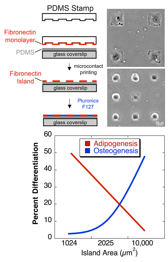
Figure 2. Control of cell shape through microcontact printing controls differentiation of MSCs.
(top) Polydimethylsiloxane (PDMS) stamps with micron-sized features are coated with fibronectin, or other ECM proteins. Fibronectin is transferred from the raised features on the stamp to a PDMS-coated glass coverslip substrate via microcontact printing. Gaps between fibronectin islands are passivated to prevent cell adhesion by adsorption of the non-adhesive, Pluronic F127. By controlling the size of the islands where cells can attach, their shape can be predefined. Phase images (courtesy of R. Desai) of cells patterned on 50×50 or 25×25 µm2 islands. (bottom) human MSCs that were allowed to adhere, flatten, and spread underwent osteogenesis, while unspread, round cells underwent adipogenesis. This switch in lineage commitment was regulated by cell shape through the modulation of endogenous RhoA activity [Data from (McBeath et al., 2004)].

