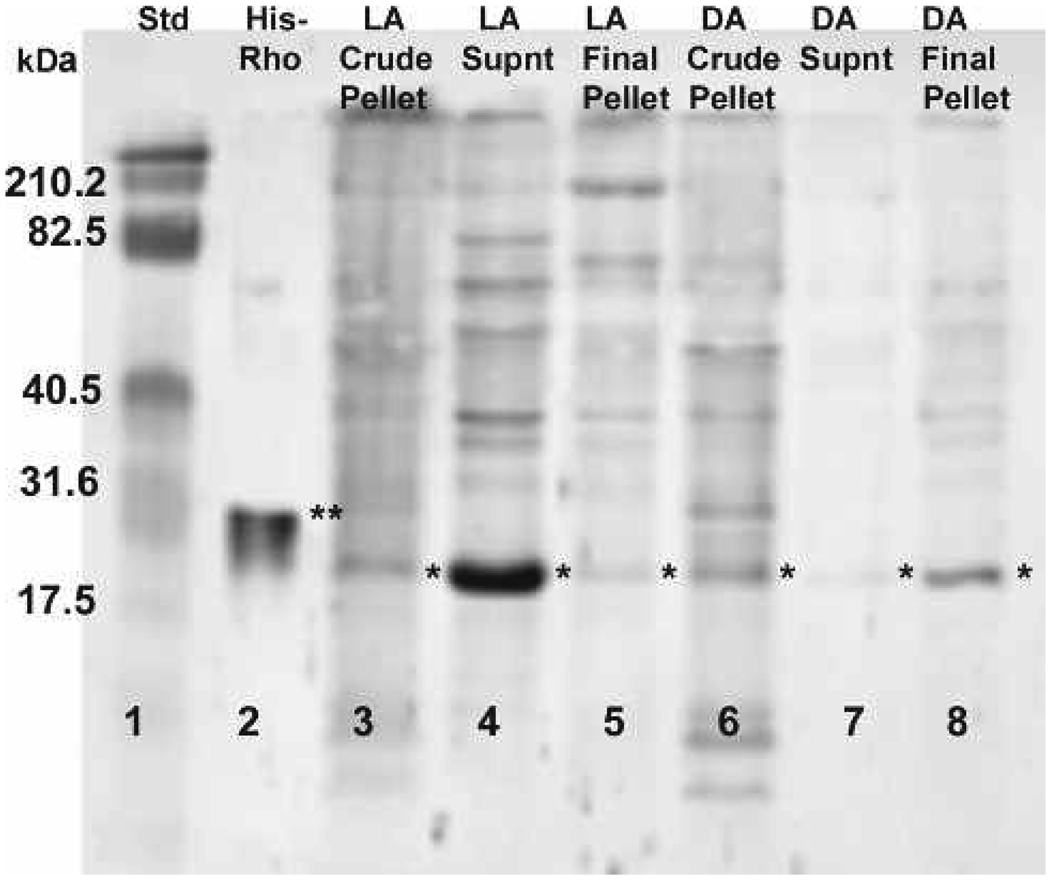
Figure 2.
Western blot localizing Rho in purified light- and dark-adapted rhabdom membrane and supernatant fractions. Rho (⋆) is present in dark-adapted (DA) rhabdom membrane fractions (lanes 6 and 8) but not in the supernatant (lane 7). In the light-adapted (LA) supernatant fraction (lane 4), Rho is present while there is little or no detection of Rho in the LA rhabdom membrane fractions (lanes 3 and 5). The double asterisk denotes the His-tagged RhoA control protein (lane 2). The molecular weight sizes correspond to the fragments of the Kaleidoscope pre-stained standard (lane 1).

