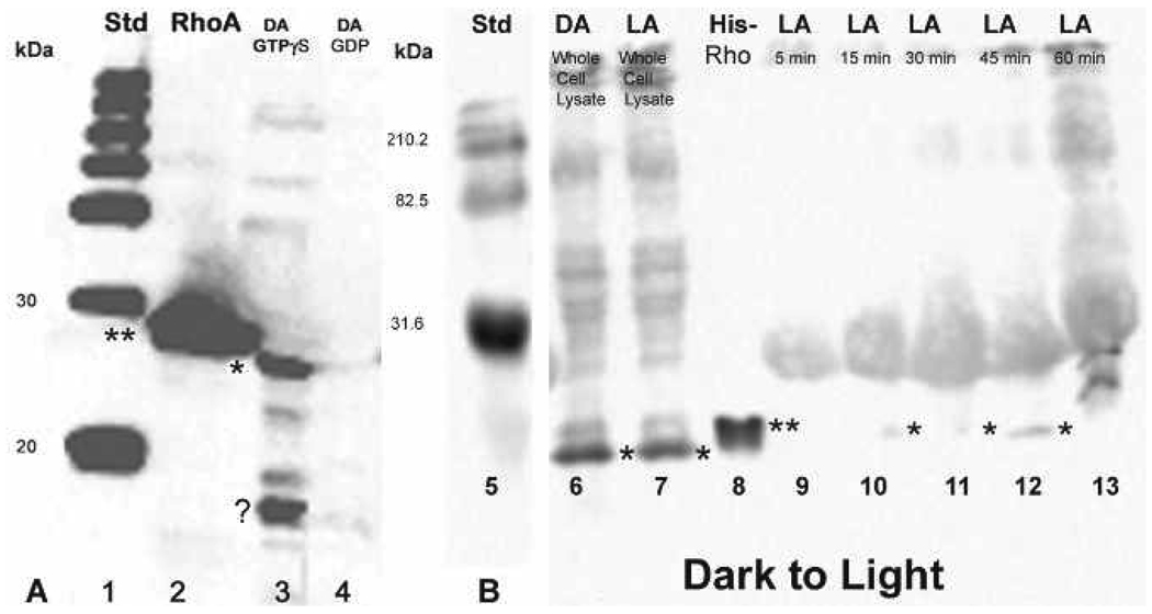
Figure 6.
A, Western blot showing controls for the Rho pull-down assay for octopuses dark-adapted and then moved to the light (lanes 1–4). There is no activation in the dark-adapted GDP control (lane 4), but there is a strong band in the GTPγS control (lane 3, ⋆⋆). The strong band migrating below 20 kDa, is also present (⋆). Lane 2 contains the His-tagged RhoA control protein and standards are in lane 1. B, Western blot after Rho pull-down assay showing the detection of weak, residual Rho activation in dark-adapted retinas moved to the light and sampled at 5, 15, 30, 45, and 60 minutes after the shift. The detection of activated Rho (⋆) is visible at 15, 30, and 45 minutes after the shift to the light (lanes 10–12). Rho in whole cell lysates (activated and inactivated) is shown in lanes 6 and 7 and standards in lanes 1 and 5.

