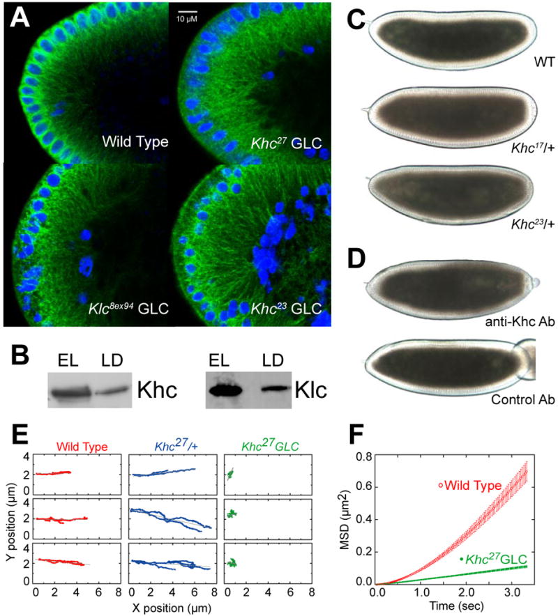
Figure 2. Kinesin-1 is directly responsible for droplet motion.

(A) Microtubule tracks are present in embryos with disrupted Kinesin-1 function. Microtubules and nuclei were visualized by staining for β tubulin (green) and DNA (blue). Radially arranged microtubules are present in the embryo periphery of all genotypes shown, though their detailed arrangement is altered in the mutants: microtubules display less apical bundling (around the nuclei) and reach deeper into the embryos; sometimes regional patches are devoid of microtubules, possibly because nuclei failed to migrate to those regions earlier in development (not shown).
(B) Both subunits of Kinesin-1 (Khc and Klc) are present on purified lipid droplets. Equal amounts of protein from total embryo lysate (EL) and biochemically purified lipid droplets (LD) were analyzed by Western blotting.
(C) Khc alleles dominantly slow net droplet motion. Embryos from Khc17 or Khc23 heterozygous mothers (i.e. carrying both a mutant and a wild-type Khc allele) often display less efficient peripheral clearing in Phase II than embryos from wild-type (top) mothers. Embryos shown are at very similar developmental stages (see movie S7 for a time-lapse comparison). This clearing defect was variable in penetrance and severity; we estimate that at least half of embryos from either genotype were distinguishable from the wild type.
(D) Acute inhibition of Kinesin-1 promotes net minus-end droplet transport. Wild-type mid-Phase II embryos were injected (on the right) with antibodies at concentrations of ∼2-3 μg/μl. Embryos injected with a function-blocking antibody against Khc (top) rapidly turned opaque near the injection site, indicating outward motion of lipid droplets. Injection of a control antibody (bottom) had no effect.
(E) In the absence of Khc, droplet motion is severely impaired. Lipid droplets were tracked in Phase II embryos; three sample traces are shown per genotype. Wild-type (WT) and Khc27/+ embryos show similar traces with bidirectional transport; Khc27 GLCs display almost no directed motion.
(F) Droplets in Khc27 GLC embryos exhibit minimal directed motion, as assessed by the Mean Squared Displacement as a function of time. The quadratic shape for wild type (WT) indicates directed transport; the reduced, linear MSD for the Khc27 GLC embryos is reminiscent of diffusion. Error bars are standard errors of the mean.
