
Figure 4.
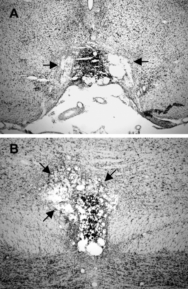
A, Representative section at −5.8 mm relative to bregma for a rat with a complete electrolytic lesion of the IPN. The black arrows show damage that extends bilaterally beyond the borders of the IPN into the paranigral nucleus. B, Representative section at −6.7 mm relative to bregma from the same electrolytic lesioned rat in A. Notice that, at this level, the lesion is complete but extends unilaterally into the ventral tegmental area.