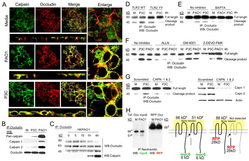
Figure 4.
Occludin is a calpain substrate. (A) Co-localization of calpain (green) and occludin (red) in polarized 16HBE cells following 1 h stimulation with heat killed PAO1 or P3C is shown in xy and xz sections. The adjacent panel shows an enlargement of white boxed area of the xy image. (B) Immunoprecipitation of occludin from 1HAEo- cell following stimulation with heat killed PAO1 or P3C and detection of occludin, pan-calpain, calpain 1 and calpain 2 by immunoblot. Data are representative of at least three separate experiments. (C) Immunoprecipitation of occludin from 1HAEo- cells following heat killed PAO1 stimulation and detection of the 80 kD hyperphosphorylated form of occludin, 60 kD full length form of occludin and 45kD cleavage fragment of occludin. (D) Detection of occludin cleavage in 1HAEo- cells overexpressing TLR2 WT or TLR2 Y616A/Y761A (TLR2YY). (E, F) Detection of occludin cleavage in cells treated with 6 μM BAPTA/AM, 20 μM ALLN, 20 μM GM6001, or 25 μM Z-DEVD-FMK. (G) Detection of occludin cleavage in P3C stimulated scrambled control or calpain 1 and 2 siRNA (CAPN 1 & 2) expressing cells. Silencing of calpain 1 and 2 expressions in scrambled control and siRNA expressing cells is shown in the adjacent panel. (H) Neutravidin immunoprecipitation (IP) from biotinylated 1HAEo- cells that were transfected (Txf) with C-terminal myc6 tagged occludin (Occ myc6) or N-terminal RFP tagged occludin (RFP Occ) and incubated with media alone (M) or heat killed PAO1 (PAO1) were immunoblotted (WB) with anti-myc or anti-RFP antibodies. Cartoon illustrates the forms of occludin that correspond with the bands on the immunoblot. (Data are representative of at least three separate experiments).

