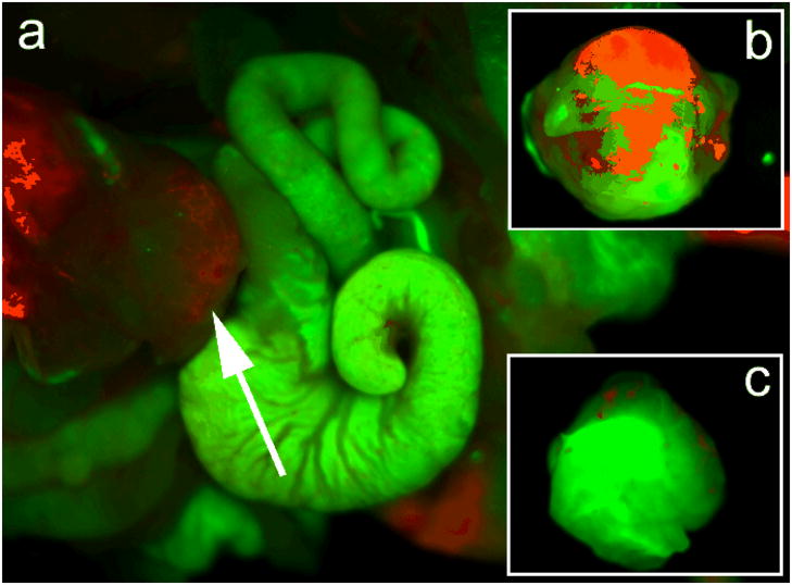
Figure 8.
(a) In vivo luminescence imaging of orthotopic tumor model mouse (tumor is pointed by white arrow) injected with ~1 mg of cRGD-peptide conjugated QRs. The autofluorescence from mice is coded green color and the unmixed QR signal is coded red color. (b) Ex vivo orthotopic tumor luminescent images of unconjugated QRs and (c) cRGD conjugated QRs harvested from mice at 2 hours, postinjection. The autofluorescence from tumor is coded green color and the unmixed QR signal is coded red color.

