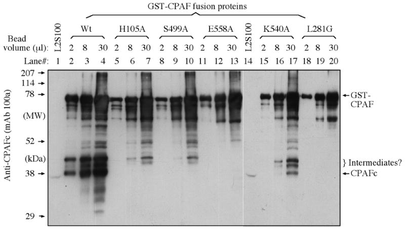
Figure 5.

Detection of CPAF processing using a Western blot. The various GST-CPAF fusion proteins were loaded onto a SDS PAGE gel as described in the legend to Figure 3. After electrophoresis, the proteins bands were transferred onto nitrocellulose membrane for detection with the anti-CPAFc mAb 100a. All CPAF preps displayed the full-length GST-CPAF fusion protein along with fragments or varying length. However, only the Wt (lanes 2-4) and the unrelated CPAF mutant K540A (lane 17) displayed a protein band migrating at the position similar to band of CPAFc from the L2S100 sample (lanes 1 & 14).
