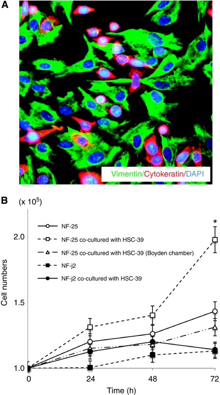
Figure 1.
Cell–cell contact with SGC-derived HSC-39 cells upregulated NF-25 gastric fibroblasts’ growth. (A) Immunofluorescence of NF-25 fibroblasts co-cultured with HSC-39 cells. NF-25 fibroblasts and HSC-39 cells were stained with vimentin (green) and cytokeratin (red) ( × 200). (B) Growth curves of NF-25 gastric fibroblasts and NF-j2 intestine fibroblasts in the presence or absence of co-incubation with HSC-39 cells. Before counting of the numbers of NF-25 and NF-j2 fibroblasts, HSC-39 cells co-cultured were excluded by separation by a magnetic beads method. To examine the effect of soluble factors, NF-25 fibroblasts and HSC-39 cells were separately co-maintained using a 1-μm pore-sized Boyden Chamber inserts. *<0.01.

