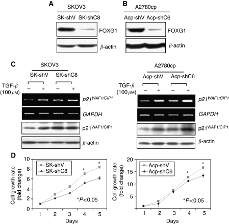
Figure 6.
Depletion of FOXG1 sensitises TGF-β mediated of p21WAF1/CIP1 induction and cell growth inhibition. (A, B) Western blot analysis showed the reduction of endogenous FOXG1 by FOXG1 shRNAi plasmid, pTER-shFOXG1, SKOV3 and A2780cp ovarian cancer cell lines, respectively. SK-shV and Acp-shV are their corresponding vector controls. (C) The vector controls and FOXG1 stable knockdown clones of SKOV3 (SK-shV and SK-shC8) (left), and A2780cp (Acp-shV and Acp-shC6) (right) were cultured in the presence (+) or absence (−) of 100 pM TGF-β for 24 h. The p21WAF1/CIP1 mRNA and protein levels were analysed by semi-quantitative RT–PCR and western blot analyses, respectively. (D) FOXG1 knockdown clones of SKOV3 (SK-shV and SK-shC8) (left), and A2780cp (Acp-shV and Acp-shC6), cultured in the medium supplemented with 10 pM TGF-β for 5 days. XTT assay shown higher cell proliferation rate in FOXG1-depleted clones (26% for SK-shC8 and 20% for Acp-shC6, P<0.05) as compared with their vector controls.

