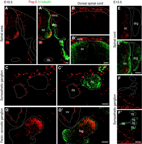
Figure 3.
Friend-of-GATA (Fog)-2 protein detection in the neural crest and its derivatives at E10.5 and E13.5. (A–D) E10.5 and (E–F) E13.5. Each panel is represented as single and double-stained slide: (A–F) Fog-2 immunohistochemistry (red), (A′–F′) double immunohistochemistry of Fog-2 (red) with β-tubulin (green) to visualise neuronal projections. (A, A′) The spinal area. (B, B′) The dorsal spinal cord with migratory neural crest cells. (C, C′) The sympathetic ganglion. (D, D′) The facio-acoustic ganglion. (E, E′) The spinal cord and dorsal root ganglion. (F, F′) Sympathetic ganglia. da: dorsal aorta; drg: dorsal root ganglion; fag: facio-acoustic ganglion; mnc: migratory neural crest; n: notochord; ov: otic vesicle; sc: spinal cord; sg: sympathetic ganglion. Bars equal 100 μm.

