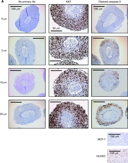
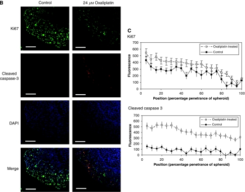
Figure 2.
The effect of oxaliplatin on proliferation and apoptosis in spheroids. Spheroids were treated with oxaliplatin (0–25 μM) for 24 h before fixation, processing and immunohistochemical assessment of Ki67 or cleaved caspase-3 (CC3) (A). Specificity of CC3 staining was confirmed using positive and negative cell pellets. The staining shown is representative of three separate experiments. Scale bars indicate either 100 μm (controls and CC3) or 50 μm (Ki67). Studies were repeated using immunofluorescence (B), and localisation of biomarkers was confirmed by the assessment of fluorescent intensity across spheroid sections (C). Sections were stained for Ki67 (green) and CC3 (red) with DAPI (blue) counterstaining to identify nuclei (B). Scale bars=50 μm. The fluorescent profile in each channel was plotted from the necrotic core to the outer rim and compared between treated and control sections to correlate staining with positional information (C). Plots are the mean intensity of 14 spheroids for each condition.


