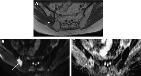
Figure 4.
A male patient with prostate cancer metastases to bone. T1W axial MRI pelvis (A) shows a metastasis within the right iliac bone (arrow). High signal within the lesion on the diffusion-weighted MRI of the pelvis (B) indicates that diffusion within the metastasis is less restricted than diffusion in the surrounding normal marrow. An apparent diffusion coefficient (ADC) map of pelvis (C) generated from the diffusion-weighted imaging data (B values 0, 50, 100, 250 500, and 750) provides a quantitative index of water diffusion within the tumour. The ADC map also shows heterogeneity of water diffusion within the tumour not shown by conventional T1W imaging.

