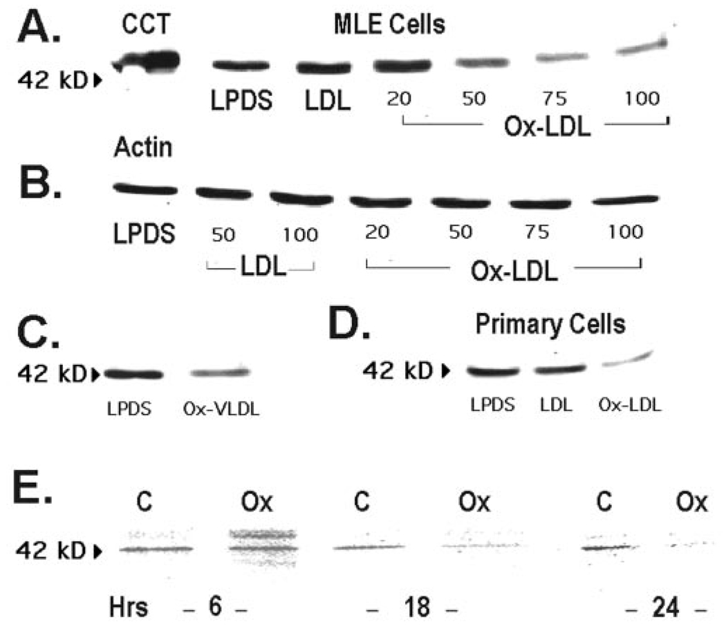
FIG. 3. Oxidized lipoproteins degrade CCTα protein.
A, MLE cells were cultured in the presence of LPDS, LPDS with LDL (100 µg/ml), or various amounts of Ox-LDL (20–100 µg/ml) for 48 h, and CCTα protein levels were determined. The leftmost lane is the purified CCTα standard (2 µg). B, levels of β-actin were determined under conditions similar to those described in A. C, cells were incubated with LPDS or in combination with Ox-VLDL (100 µg/ml) for 48 h, and CCTα levels were determined. D, primary rat alveolar type II epithelial cells were incubated for 24 h with LPDS, LPDS plus LDL (LDL, 75 µg/ml), or LPDS plus Ox-LDL (Ox-LDL, 75 µg/ml), and CCTα protein levels were determined. All lanes in panels A–D contain equal amounts of total cellular protein. E, CCTα protein degradation in MLE cells was determined by pulsing cells with [35S]methionine for 4 h; cells were rinsed and incubated with chase medium (10) for 6–24 h with or without 100 µg/ml Ox-LDL (Ox). Equal amounts of enzyme protein were immunoprecipitated with CCTα antibody. A–D, n = 4 separate studies; E, n = 2 studies.

