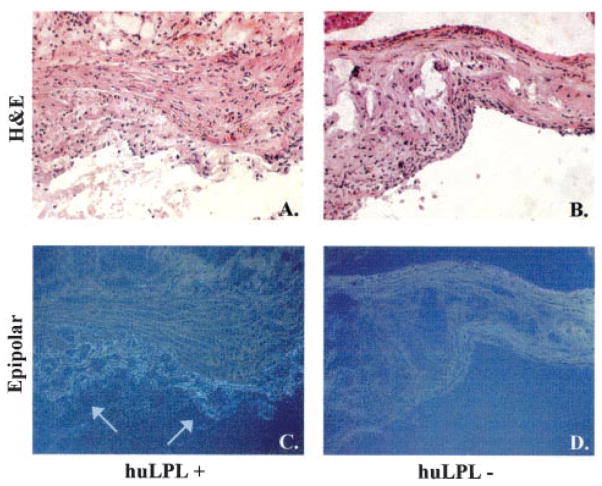
Figure 5.
huLPL protein is present in aortic lesions of transgenic mice. Male apoE KO mice with transgenic huLPL+ (A and C) or huLPL− (B and D) genotype were maintained on a Western diet for 17 weeks. Aortic sections were processed for immunohisto-chemistry with an huLPL-specific monoclonal IgG 5D2 as the primary antibody. The secondary antibody used was goat anti-mouse ultrasmall gold. Images were obtained under epipolarized light (C and D). Arrows indicate staining for LPL. The same sections were also stained with hematoxylin and eosin for visualization of lesion morphology under the light microscope (A and B).

