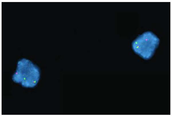
Figure 1. Breakapart FISH analysis of interphase cells from a patient with de novo RAEB-I, demonstrating rearrangement of MLL.
Red probe corresponds to the 5′ end of MLL, whereas the green probe corresponds to the 3′ end of MLL. Intact MLL is associated with an overlapping red/green (i.e., yellow) fusion signal. In this case, each cell demonstrates separation of one set of the red and green probes, corresponding to a translocation involving MLL.

