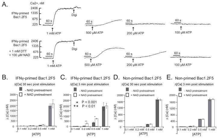
Figure 7. NAD potentiates ATP-induced P2X7R activation in interferon-primed murine macrophages that express ART2.1.
Bac1.2F5 macrophages were transferred to M-CSF-free medium and then stimulated with IFN-γ (100 U/ml) for 24 hr (Panels A-C) or were incubated in M-CSF-free medium 24 hr in the absence of IFN-γ. Cytosolic Ca2+ was measured in fura-2 loaded Bac1 cell suspensions as previously described but with inclusion of 1 mM DTT and 1 mM ADP-R in all test media. The cell suspensions were pre-treated with a cocktail of 50 μM ADP and 50 μM UTP to activate and desensitize P2Y receptors 5 min prior to P2X7R stimulation by the indicated concentrations of ATP; where indicated (as in the panel B traces) 100 μM NAD was also added 5 min prior addition of ATP. In each assay, the cells were permeabilized with digitonin (digi) for calibration of Ca2+− dependent fura-2 fluorescence. A) ATP-induced Ca2+ influx without or with NAD pre-treatment for 5 min. These traces are representative of observations from 4 experiments. B, C) Changes in cytosolic Ca2+ in IFN-γ-primed Bac1 macrophages were quantified at 30 s (panel B) or 3 min (panel C) after addition of the indicated concentration of ATP with or without NAD pretreatment. Data bars represent the mean±SE from 4 experiments. D, E) Identical experiments as in panels B and C, but with Bac1 macrophages that were not primed with IFN-γ. Data bars represent the mean±SE from 4 experiments.

