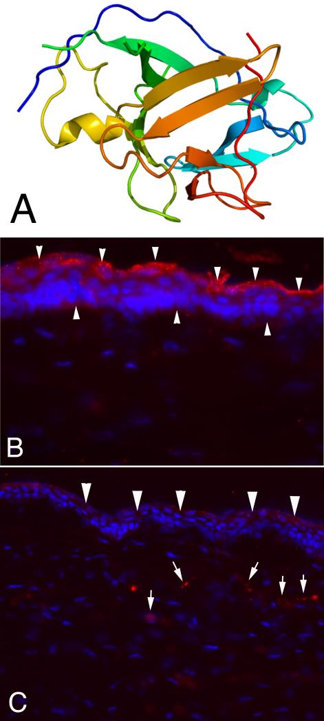
Fig.
Model of structure of IL-1α and expression in unwounded and wounded rabbit corneas. A. Model structure of human interleukin-1 alpha. B. IL-1α is expressed constitutively throughout the corneal epithelium (arrowheads, red), but appears to be most highly expressed in apical epithelial cells. Note that little IL-1α is associated with keratocyte cells in the stroma of the unwounded cornea. Mag. 500X C. At one month after injury (in this case -9 diopter PRK that triggers haze and myofibroblast development) IL-1α is now detected in stromal cells (arrows, red), in addition to continued expression in the healed epithelium (arrowheads, red). Blue stain is DAPI staining of cell nuclei. Mag. 500X.

