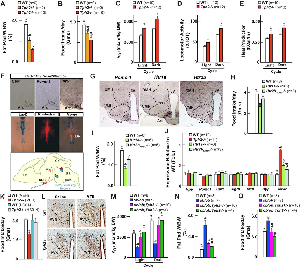
Figure 6. Serotonin promotes food intake through Htr1a and Htr2b receptors on arcuate neurons.
(A–B) Fat pad weights (A) and food intake (B) in WT, Tph2+/− and Tph2−/− mice.
(C–E) Energy expenditure in WT and Tph2−/− mice; measured by volume of oxygen consumption (VO2) (C), locomotor activity (D) and Heat production (E).
(F) Analysis of axonal projections emanating from the serotonergic neurons. Cross of Sert-Cre and Rosa26REcfp mice identified projections reaching arcuate (Arc) nuclei in the hypothalamus through Ecfp immunohistochemistry colocalized to molecular markers of arcuate neurons (Pomc-1 and Npy) by in situ hybridization. Retrograde Rhodamine dextran labeling of the arcuate neurons identified serotonergic neurons in the brainstem in Tph2LacZ/+ mice through colocalization of β-galactosidase staining and Rh-dextran fluorescence in serotonergic neurons of the brainstem.
(G) In situ hybridization analysis of Htr1a, Htr2b in Pomc1-expressing arcuate neurons of the hypothalamus. 3V: third ventricle.
(H–I) Food intake (H) and fat pad weights (I) in WT, Htr1a−/− and Htr2bPOMC−/− mice.
(J) qPCR analysis of hypothalamic gene expression in WT, Htr1a−/− and Htr2bPOMC−/− mice.
(K) Food intake in WT, Tph2−/− mice before and after Mc4r antagonist (HS014) administration.
(L) cFos induction in paraventricular nucleus of hypothalamus in WT, Tph2−/− mice before and after acute administration Mc4r agonist (MTII). 3V: third ventricle.
(M–O) Volume of oxygen consumption (M), fat pad weight (N) and food intake (O) in WT, ob/ob, ob/ob;Tph2+/− and ob/ob;Tph2−/− mice.
All panels (except A–B, H–J and M–O) * P < 0.05; ** P < 0.01 (Student’s t test). Error bars, SEM. Panels A–B, H–J and M–O (One way ANOVA, Newman-Keuls test); Different letters on 2 or more bars indicate significant differences between the respective groups (P < 0.05).

