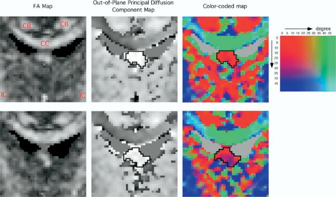
Figure 2.
Fractional anisotropy map, out-of-plane principal diffusion component map, and color-coded map. Upper row: the selected slice. Lower row: the slice just anterior to the selected slice. The boundary of fornix is shown in the out-of-plane principal diffusion component maps and the color-coded maps. In out-of-plane principal diffusion component map, high-intensity corresponds to the fiber tract perpendicular to the plane, and the fornix is easy to differentiate. Two angle maps were merged in this color-coded map to make it more intuitively understandable to the reader. One is a map for the angle between the first eigenvector and the sagittal plane. The other is a map for the angle between the first eigenvector and the axial plane. The color table shows these two angular values of each color. Red color means the first eigenvector closely perpendicular to the coronal plane. Green color means the eigenvector with medial-lateral direction. Green color in the superior part of each color-coded image corresponds to the corpus callosum. Blue color means the eigenvector with superior–inferior direction. Blue color in the inferior part of both sides corresponds to the internal capsule. Blue color in the delineated fornix of the slice just anterior to the selected slice indicates that the fornix is descending inferiorly in this slice. CB, cingulum bundle; CC, corpus callosum; IC, internal capsule.

