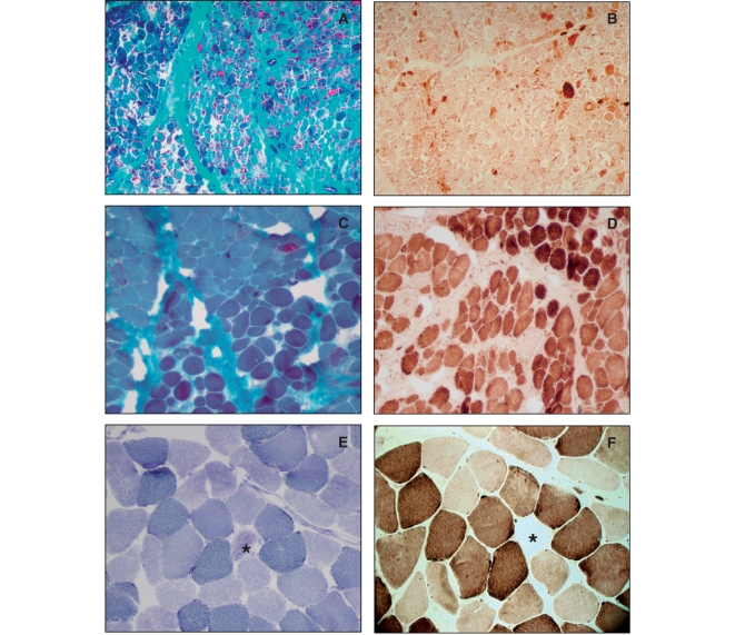
Figure 2.
Histochemical staining of the muscle biopsy from Patient 7 at 3 months of age (A and B) and 9 years (C and D) and of the muscle from his asymptomatic mother (E and F). The early biopsy confirmed severe mitochondrial myopathy with RRF (A) and even more COX-negative fibres (B). These changes significantly improved but did not completely disappear at 9 years of age (C and D). The muscle biopsy from the mother revealed a few COX-negative (F), SDH hyper-reactive (E) fibres (stars). A, C: Gomori trichrome; B, D, F: COX; E: SDH stain. Magnification: A, B 100×, C, D, E, F 200×.

