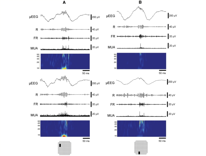
Figure 5.
HFOs occurring simultaneously during a macrodischarge recorded from Patient 4. Two pairs of adjacent channels in different areas of the MEA are shown (A, B; solid black boxes in schematics). The same time slice is shown in all four panels, with signals displayed as in Fig. 1. Note the marked differences in timing, morphology, and frequency range in both the 100–500 Hz bands and in MUA between adjacent recording sites. For example, in (B) the maxima of the fast ripple amplitude envelope are clearly seen to occur at different times in the two channels, while in (A) there is fast ripple activity in the top channel that is not reflected in the bottom channel. These observations indicate that recording sites separated by 400 μm are capable of distinguishing independent generators of high frequency activity.

