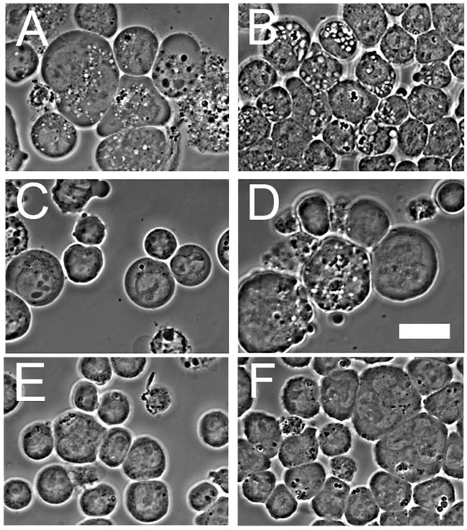
Fig. 6.
Phase-contrast images showing development of autophagic vacuoles. (A) = wild-type L1210 cells 15 min after exposure to HA14-1 (30 µM); (B) = L1210/Bax− under similar conditions; (C) = L1210 cells 30 min after an LD90 PDT dose with CPO; (D) = L1210/Bax− under similar conditions; (E)=control L1210; (F)=control L1210/Bax−.White bar in panel (D) = 10 µm.

