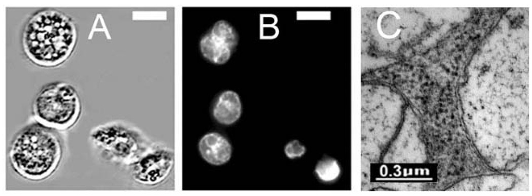
Fig. 7.
(A) = Autophagic vacuole formation in L1210/Bax− cells after an LD90 dose of PDT (CPO); (B) = HO33342 labeling of nuclei showing lack of apoptotic fragmentation; (C) = electron microscopic examination of vacuoles seen in Fig. 5(A) showing the double membrane characteristic of autophagy. White bars in panels (A) and (B) = 10 µm.

