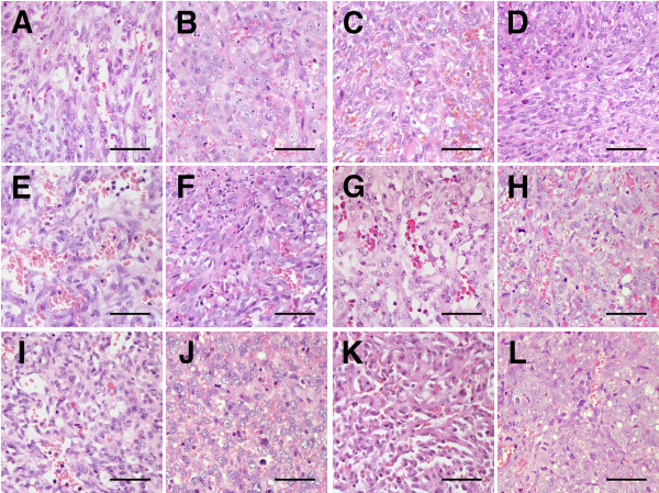
Figure 2.
Histological features of original HSAs and xenograft HSA tumors. Microscopic features of original HSAs and xenograft HSA tumors. Histologically, each of the original HSAs (A, C, E, G, I, and K present the original tumors of Si, Re, Ud, Sy, Sa, and Ju, respectively) showed vascular-like proliferation and formed solid sheets with various-sized clefts. The neoplastic cells that comprised original tumors had various shapes, ranging from spindle-shaped and polygonal to ovoid. Each of the xenograft tumors (B, D, F, H, J, and L present Si, Re, Ud, Sy, Sa, and Ju, respectively) were predominantly solid and contained poorly formed, irregular-shaped vascular spaces with erythrocytes, neutrophils, and lymphocytes. In all xenograft tumors, the neoplastic cells became more pleomorphic, elongated, and plump and the nuclei became larger and more clearly polygonal in shape. Hematoxylin and eosin (HE); bars: 50 μm.

