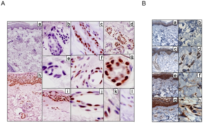
Figure 7. Elevated Nab2 expression in scleroderma skin biopsies.
Lesional skin biopsies were obtained from patients with scleroderma (n = 6) and healthy controls (n = 3), and processed for immunohistochemistry as described under Methods. A. Nab2 expression. Skin biopsies from healthy controls (a, b, e) and scleroderma patients (c, d, f, g, h–l). Original magnification ×100(a, h), ×400 (b, d, i), ×630 (c, e, f, j, k, l). Note uniformly intense Nab2 nuclear immunoreactivity in epithelial cells in scleroderma skin biopsies (h, i, l), as well as in perifollicular and periglandular (c, d, g) epithelial cells and vascular endothelial cells (f, j), compared to weak and variable Nab2 expression seen in occasional dermal fibroblasts (k, l). Representative photomicrographs; similar immunostaining pattern was observed in all six scleroderma skin biopsies. B. Phospho-Smad2 (a–d) or Egr-1 (e–h) expression. Nuclei are counterstained with hematoxylin (blue). Note strong immunostaining in dermal fibroblasts in scleroderma (d, h) skin biopsies compared to healthy controls (b, f).

