Figure 6.

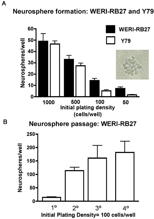
Neurosphere formation in retinoblastoma cultures. A: Y79 and WERI-R27 cells were plated as single cell suspensions in 96 well dishes at initial plating densities of 50-1,000 cells per well, in triplicate. Low cell densities were chosen to minimize effects of non-specific cell aggregation in favor of neurospheres originating from one single cell. After five days, neurospheres were counted and results presented. Both Y79 and WERI-RB27 human retinoblastoma cells formed neurospheres at all cell densities tested. A typical neurosphere (WERI-RB27) is shown (inset). B: WERI-RB27 cells were prepared as single cell suspensions at an initial plating density of 100 cells per well, in triplicate. Every 3-4 days, neurospheres were counted and then dissociated into single cell suspensions, diluted 1:2, and replated. The graph depicts the number of neurospheres counted at each passage.
