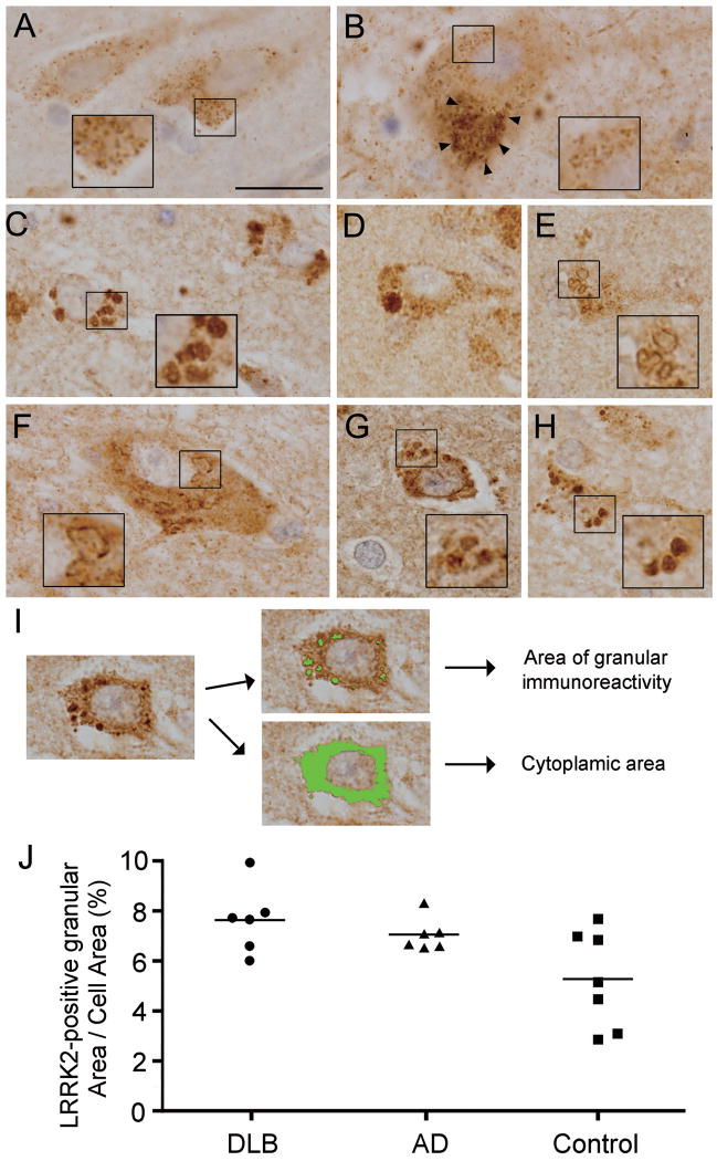
Figure 4.
Leucine-rich repeat kinase 2 (LRRK2)-positive enlarged granules and vacuoles. (A, B) In normal aged controls, LRRK2 immunoreactivity has a small punctate pattern throughout the neuronal soma and processes in the entorhinal cortex (A) or neuromelanin-containing dopaminergic neurons (arrowheads) of the substantia nigra pars compacta. (C-E) In the entorhinal cortex of DLB cases, LRRK2 immunoreactivity in neurons is mostly in abnormal enlarged (C) or giant (D) granules or clustered vacuoles (E). (F, G) LRRK2-positive vacuoles or granules in neurons of the substantia nigra pars compacta (F) and entorhinal cortex (G) from PD cases. (H) LRRK2-positive enlarged granules in neurons of the entorhinal cortex from an AD case. Scale bar in (A) represents 20 μm in A-H. (I) Computerized image analysis for estimation of the volume of enlarged granules immunostained with anti-LRRK2 antibody (JH5514) or total cytoplasmic area outlined by brown (DAB) immunoreactivity, as described in Methods. (J) Percentage of total cell area occupied by LRRK2-positive granules in neurons of the entorhinal cortex of DLB brains (n = 6), AD brains (n = 6) and aged control brains (n = 7). Values are the mean percentage calculated from 25 neurons per brain.

