
Fig. 2.
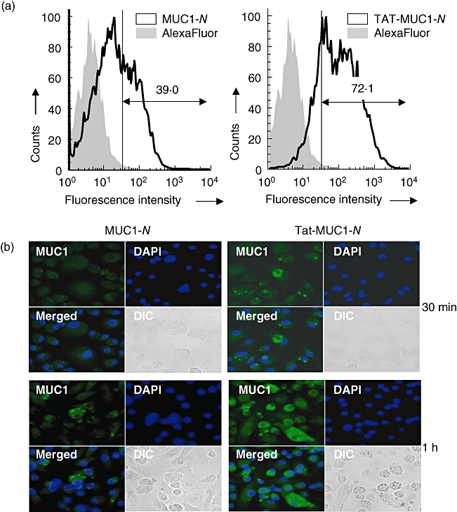
Transduction of recombinant N-terminal region mucin antigen 1 (MUC1) into dendritic cells (DCs). (a) Flow cytometric analysis of MUC1 in DCs transduced with N-terminal region MUC1 (MUC1-N) or Tat-MUC1-N. DCs were incubated with either free AlexaFluor 594 (grey area) or AlexaFluor 594-labelled proteins (solid line) for 30 min. (b) Confocal microscopic analysis of DCs transduced with MUC1-N or Tat-MUC1-N. After incubation of DCs with recombinant MUC1 proteins for 30 min or 1 h, intracellular MUC1 was stained with anti-human MUC1-fluorescein isothiocyanate (green). Nuclei were stained with 4,6-diamidino-2-phenylindole (DAPI) (blue). Two pseudocoloured images (MUC1 + DAPI) were merged (merged), and differential interference contrast (DIC) images are shown.