
Fig. 3.
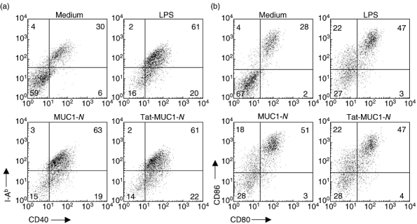
Expression of co-stimulatory molecules and I-Ab of dendritic cells (DCs) pulsed with recombinant mucin antigen 1 (MUC1). Because it has been known that MUC1 could impair DC function, we analysed co-stimulatory molecule expression on DCs after they were pulsed with purified N-terminal region MUC1 (MUC1-N) or Tat-MUC1-N proteins. After DCs were incubated with lipopolysaccharide for another 18 h after protein pulsing, cells were stained with antibodies reactive with I-Ab, CD40, CD86, CD80 and CD11c. Live DCs (7AADneg/CD11cpos) were analysed for the surface molecule expression using a flow cytometer.