
Fig. 5.
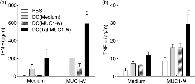
Interferon (IFN)-γ (a) and tumour necrosis factor (TNF)-α (b) secretion of lymph node (LN) cells in response to mucin antigen 1 (MUC1). LN cells were harvested as in Fig. 4 and cultured in the presence [N-terminal region MUC1 (MUC1-N), 50 µg/ml] or absence of MUC1-N proteins (medium) for 48 h. The levels of cytokines in the culture supernatants were measured by cytometric bead array. Each sample was analysed in triplicate. Results are presented as mean ± standard error of the mean. *P < 0·05 or #P = 0·08 when compared with DC (MUC1-N).