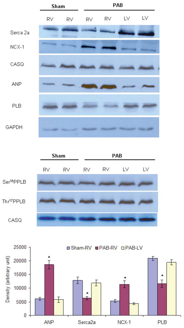
Figure 2.

Protein Quantitation in the ventricular tissues of sham-operated and PAB-rabbits. (A, B) Different SDS-PAGE gel concentrations (10% for Seca2a and CASQ, 9% for NCX-1, 15% for ANP, 14% for PLB, Ser16PPLB and Thr17PPLB) were used to resolve total ventricular homogenates and immunoprobed with specific antibodies. A total of 12 µg (panel A) or 20 µg (panel B) of total homogenate was loaded in each well. GAPDH and CASQ were used as loading controls. (C) The expression of SERCA2a and PLB was significantly decreased while expression of ANP and NCX-1 was significantly upregulated in the right ventricle of PAB-rabbits (PAB-RV) in comparison to the left ventricle of PAB-rabbits (PAB-LV) and right ventricle of sham-operated rabbits (Sham-RV). Values given are mean ± SEM from 4 rabbits/group (unpaired t-test). * indicates values significantly (p<0.05) different in comparison to both sham-RV and PAB-LV. PAB, pulmonary artery banding; RV, right ventricle; LV, left ventricle.
