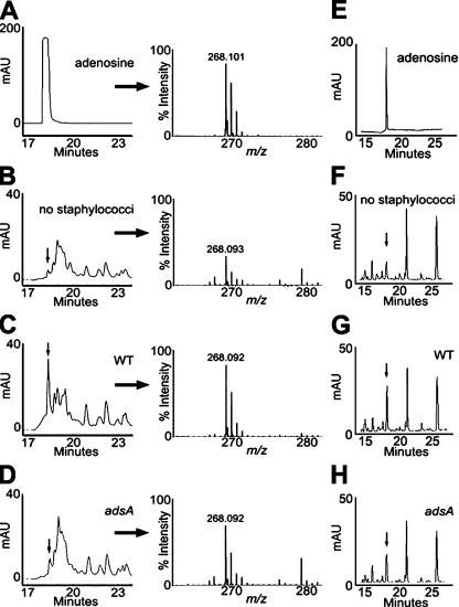
Figure 4.
S. aureus AdsA synthesizes adenosine in blood. (A) RP-HPLC to quantify 100 µM adenosine (left) and identify its monoisotopic ions by MALDI-MS (right). 1 ml of lepirudin-anticoagluated mouse blood was incubated without (B) or with 105 CFU of wild-type S. aureus Newman (WT; C) or its isogenic adsA variants (D) for 1 h. Plasma was deproteinized, filtered, and subjected to RP-HPLC to quantify adenosine (left) and identify its monoisotopic ions by MALDI-MS (right). Arrows denote corresponding adenosine peaks. Calculated abundance of adenosine in plasma extrapolated from the purified adenosine control was 1.1 µM (B, no staphylococci), 13.2 µM (C, WT S. aureus Newman), and 2.1 µM (D, adsA mutant staphylococci). Data are representative of three independent analyses. (E–H) RP-HPLC analyses of 50 µM adenosine (E) or plasma collected from mice that had been mock infected (F), or from animals that were challenged with 107 CFU wild-type (WT; G) or (H) adsA mutant bacteria. Data are representative of two independent analyses. The calculated abundance of plasma adenosine in mice extrapolated from the purified adenosine control was 2.8 ± 0.6 µM (F, no staphylococci), 16.4 ± 2.1 µM (G, WT S. aureus Newman), and 7.4 ± 3.2 µM (H, adsA mutant staphylococci). mAU, milliabsorbance units of HPLC eluate.

