Figure 5.
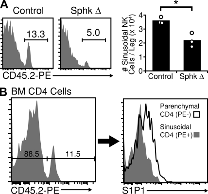
Reduced numbers of NK cells in BM sinusoids of S1P-deficient mice and S1P1 surface expression on BM T cells. (A, left) NK cells present in the BM sinusoids of Sphk-deficient and littermate control mice were labeled in vivo by a 2-min i.v. treatment with PE-conjugated anti-CD45.2. Numbers indicate the frequency of PE+ (sinusoidal) cells among total NK cells. (right) Summary of data for three mice. Data are representative of two independent experiments, each involving at least two Sphk-deficient and two littermate control mice. Bars represent mean values, and circles represent individual animals. (B, left) Control mice were treated i.v. with PE-conjugated anti-CD45.2 for 2 min. Cells were gated on CD4+, TCRβ+, NK1.1− cells and resolved for sinusoidal labeling. (right) Comparison of S1P1 surface expression levels between PE− (parenchymal) and PE+ (sinusoidal) CD4+ T cells. Data are representative of two experiments, each analyzing two animals. *, P < 0.05.

