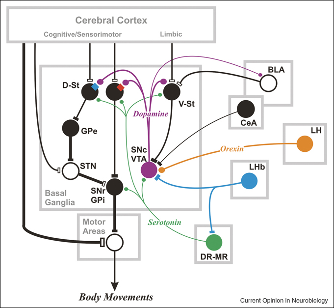
Figure 1.
Subcortical areas and their connections that can control reward-directed behavior. Central to the subcortical network is the basal ganglia which are capable of disinhibiting actions that lead to rewarding outcomes and suppressing actions that lead to non-rewarding or punishing outcomes. The basal ganglia mechanism becomes functional owing to signals mediated by neuromodulators (e.g. dopamine, serotonin, orexin, acetylcholine) and signals originating outside the basal ganglia (e.g. amygdala, lateral habenula), in addition to the inputs from the cerebral cortical areas. The target of the SNr-GPi, denoted as ‘Motor Areas’, may be subcortical motor areas, such as the superior colliculus, or ventral thalamic nuclei which are connected with motor cortical areas. Direct pathway neurons in D-St express dopamine D1 receptors (red square); indirect pathway neurons express D2 receptors (blue square). V-St may include striosomes (or patches) in the D-St. For clarity, many connections and neuron types are omitted. BLA: basalateral amygdala, CeA: central amygdala, DR: dorsal raphe, D-St: dorsal striatum, GPe: external segment of globus pallidus, GPi: internal segment of globus pallidus, LH: lateral hypothalamus, LHb: lateral habenula, MR: medial raphe, SNc: substantia nigra pars reticulata, SNr: substantia nigra pars reticulata, STN: subthalamic nucleus, V-St: ventral striatum, VTA: ventral tegmental area. Filled circles indicate inhibitory neurons; open circles indicate excitatory neurons. LHb neurons inhibit SNc-VTA and DR-MR neurons, but their action is mediated by inhibitory interneurons. SNc-VTA contains dopamine neurons, DR-MR contains serotonin neurons, and LH contains orexin neurons, all of which exert modulatory effects on their target neurons.

