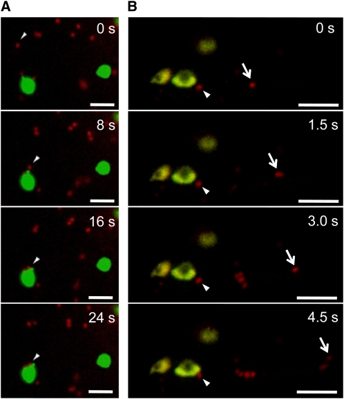
Figure 4.
Two-Dimensional Time-Lapse Imaging of a Communication between Golgi and Plastid.
ST-mRFP and WxTP-GFP dual labeling with AmyI-1 expression was performed in onion cells. ST-mRFP, red; WxTP-GFP, green. Bars = 5 μm.
(A) The images were taken at the rate of 1 frame per 4 s. Cropped frames from the movie (see Supplemental Movie 1 online) are shown here. Arrowhead shows the Golgi body (red) soft-landing on the surface of the plastid (green).
(B) The images were taken at a rate of 1 frame per 0.5 s. Cropped frames from the movie (see Supplemental Movie 2 online) are shown here. Arrows show Golgi bodies with different locomotion characteristics: one remains around the plastids (arrowhead) and the other exhibits active movement (arrow).

