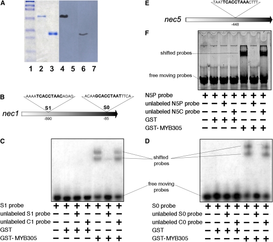
Figure 6.
MYB305 Binds to the nec1 and nec5 Promoter Fragments.
(A) Expression and purification of LxS-MYB305 protein. Lanes 1 to 3 are Coomassie blue stained: lane 1, protein standards; lane 2, purified GST-MYB305 protein; lane 3, GST protein. Lanes 4 to 7 are immunoblots using purified anti-LxS8-MYB305 antibodies: lane 4, purified GST-MYB305 protein; lane 5, GST protein; lane 6, 100 μg of protein from Stage 12 nectary tissue of wild-type LxS8 plant; lane 7, 100 μg of protein from foliage of wild-type LxS8 plant.
(B) Structure of the N. plumbaginifolia nec1 promoter showing the location and sequences of the two consensus MYB binding sites S1 and S0.
(C) Mobility shift of the radiolabeled S1 probe. Inclusion of individual components is indicated below each lane. S1 probe represents the S1 MYB binding site on the nec1 promoter. The unlabeled S1 probe is identical to the radiolabeled S1 probe and the unlabeled C1 probe differs from S1 probe with MYB consensus sequence replaced by poly A/T as indicated in Supplemental Table 1 online. Film exposure was 2 h.
(D) Mobility shift of the radiolabeled S0 probe. Inclusion of individual components is indicated below each lane. S0 probe represents the S0 MYB binding site of the nec1 promoter. The unlabeled S0 probe is identical to the radiolabeled S0 probe, and the unlabeled C0 probe differs from S1 probe with MYB consensus sequence replaced by poly A/T as indicated in Supplemental Table 1 online. Film exposure was 2 h.
(E) Structure of the N. plumbaginifolia nec5 promoter showing the location of the consensus MYB binding site.
(F) Mobility shift of the fluorescently labeled N5P probe. Inclusion of individual components is indicated below each lane. The unlabeled N5P probe is identical to the fluorescently labeled N5P probe, and the unlabeled N5C probe differs from N5P probe as indicated in Supplemental Table 1 online.
[See online article for color version of this figure.]

