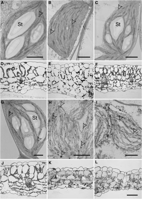
Figure 7.
Micrographs of Wild-Type and pxa1 Leaf Tissue at the End of the Light Period and after Extended Darkness.
Electron ([A] to [C] and [G] to [I]) and bright-field ([D] to [F] and [J] to [L]) micrographs of chloroplasts and leaf cross sections prepared from Col-0 wild-type ([A] to [F]) and pxa1-2 ([G] to [L]) leaves at the end of the regular light period (1st column), after 36 h of darkness (2nd column) and after 36 h of darkness plus 4 h of daylight (3rd column). Note the disintegrated structure and increased number of plastoglobules/lipid droplets in chloroplasts of dark-treated pxa1-2 leaves. St, starch granules. Arrowheads indicate plastoglobules. Bars = 1 μm in (A) to (C) and (G) to (I) and 50 μm in (L) (same scale for [D] to [E] and [J] to [L]), respectively.

