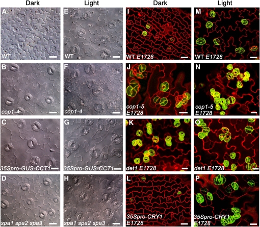
Figure 3.
COP1 Acts Constitutively to Suppress Stomatal Development and Differentiation and Is Regulated by CRY1, SPA, and DET1.
(A) to (D) DIC images of the abaxial cotyledon epidermis of 10-d-old dark-grown wild-type (A), cop1-4 (B), 35Spro–GUS–CCT1 (C), and spa1 spa2 spa3 (D) seedlings.
(E) to (H) DIC images of the abaxial cotyledon epidermis of wild-type (E), cop1-4 (F), 35Spro–GUS–CCT1 (G), and spa1 spa2 spa3 (H) seedlings grown under white light (150 μmol·m−2·s−1) for 10 d.
(I) to (L) Confocal images of the abaxial cotyledon epidermis of 7-d-old dark-grown wild-type expressing E1728 (WT E1728) (I), cop1-5 mutant expressing E1728 (cop1-5 E1728) (J), det1 mutant expressing E1728 (det1 E1728) (K), and 35Spro–CRY1 expressing E1728 (35Spro–CRY1 E1728) (L).
(M) to (P) Confocal images of the abaxial cotyledon epidermis of 7-d-old light-grown WT E1728 (M), cop1-5 E1728 (N), det1 E1728 (O), and 35Spro–CRY1 E1728 (P). Light condition for (M) to (O) is white light (150 μmol·m−2·s−1), and for (P) is blue light (50 μmol·m−2·s−1). In (I) to (P), epidermal cell periphery is highlighted by propidium iodide (PI; red), and mature guard cells are indicated by the E1728 marker (green). Bars = 20 μm.

