Fig. (3).
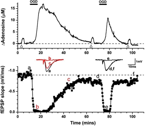
Repeated in vitro ischemia results in reduced adenosine release in hippocampal slices. Upper trace - adenosine release as measured by a MK I sensor in response to two sequential periods of oxygen/glucose deprivation (OGD; black bars). Bottom trace - depression and recovery of synaptic transmission in response to the OGD episodes. Note the reduced release of adenosine and reduced effects on the fEPSP during the second period. Inset are individual fEPSPs taken at the times indicated. Triangles refer to applications of exogenous adenosine to test that the sensor has not run down over this period.

