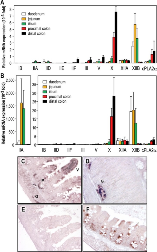
Fig. 1.
Expression of the different mouse sPLA2s in the intestine of C57BL/6J and BALB/c mice by RT-qPCR and in situ hybridization of mGX sPLA2. A and B, RT-qPCR in C57BL/6J (A) and BALB/c (B) mouse intestine sections using specific sets of PLA2 primers. To facilitate the comparison of expression between the different sPLA2s, the data were first normalized to glyceraldehyde-3-phosphate dehydrogenase mRNA, which was used as a reference gene, and then expressed relative to the lowest expression level that can be accurately measured in our RT-qPCR assay conditions [i.e., the expression of pancreatic group IB sPLA2 in the colon {relative abundance of 1 (arbitrary unit = 1)}]. Note that two different ordinate axes have been used in B. mGIIE sPLA2 could not be detected in all intestine sections from BALB/c mice (C–F). In situ hybridization of mGX sPLA2 in small intestine (ileum) showing labeling of columnar epithelial cells in mucosal villi (V), Paneth cells (P), and ganglion cells (G) of the myenteric plexus. Hybridized with antisense probe. D, absence of reaction product from ileal tissue when hybridized with sense probe. E, in situ hybridization of mGX sPLA2 in large intestine (cecum) showing labeling in epithelial cells. Hybridized with antisense probe.

