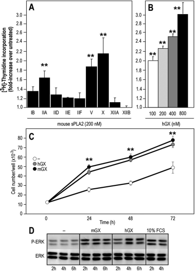
Fig. 3.
Effect of various mouse sPLA2s and hGX sPLA2 on DNA synthesis, cell growth, and p42/44 MAP kinase phosphorylation in Colon-26 cells. A and B, quiescent cells were incubated for 24 h with the different mouse sPLA2s (A) or various concentrations of hGX sPLA2 (B). [3H]Thymidine incorporation is expressed as -fold increase over untreated cells (incorporation of [3H]thymidine under untreated conditions was 44,962 dpm). Values represent average values of at least three experiments performed in triplicate (A) or are representative of at least three experiments (B). Group IIA, V, and X sPLA2s significantly stimulated [3H]thymidine incorporation compared with untreated cells (**, P < 0.001; one-way ANOVA, with Bonferroni adjustment). No significant difference was found between cells treated with group V and X sPLA2s (P = 0.2176; Student's t test). C, quiescent Colon-26 cells were cultured for up to 3 days in the presence or absence of hGX or mGX sPLA2s (200 nM) and counted every day. The difference in cell number between cells cultured in the absence and presence of sPLA2 at different time points is statistically significant (**, P < 0.05; Student's t test). D, cells were incubated for various times at 37°C with mGX and hGX sPLA2s (200 nM). The cell lysates were subjected to immunoblotting with anti-phospho-specific antibodies that specifically recognize tyrosine-phosphorylated p42/44 MAPK. Equal amounts of proteins were loaded, which was verified by immunoblotting of total p42/44 MAPK proteins.

