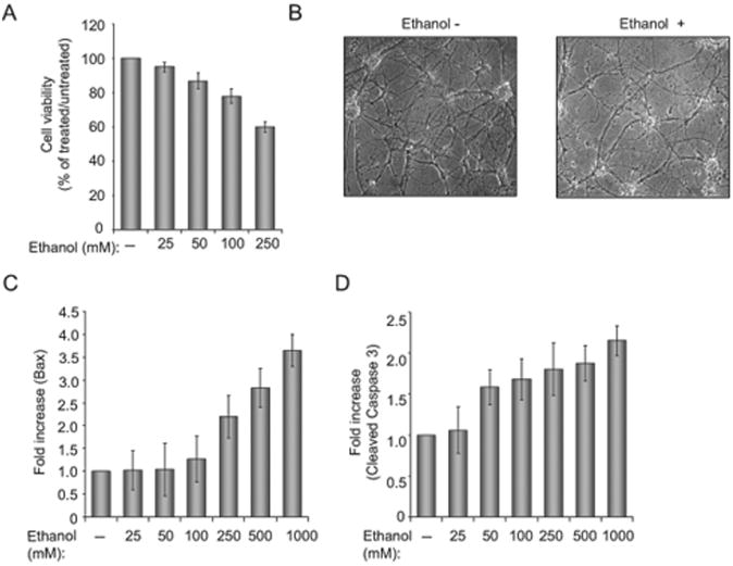
Figure 1. Effect of ethanol on neuronal cell morphology and apoptotic pathways.
(A) Cell viability assay in rat primary neurons, incubated with increasing concentrations of ethanol (from 25 to 250 mM). Equal numbers of cells were plated in duplicate, and then incubated with ethanol. Cell viability was evaluated by Trypan blue exclusion assay. Bar 1 represents untreated cells set as 100%. (B)Phase images (magnification 200×) of neuronal cells incubated in the absenceand presence of 250 mM ethanol. (C)Quantification of the differences in Bax levels as determined by densitometric analysis of the band corresponding to Bax (as determined by Western blot assay) that was normalized to the level of Grb2. (D) Quantification of the differences in cleaved caspase 3 after being normalized to the level of Grb2.

