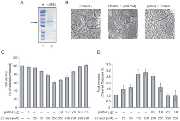
Figure 2. Effect of p38SJ on neuronal cell injury by ethanol.
(A)SDS-PAGE illustrating highly purified p38SJ that was obtained upon size fractionation of crude protein extracts from the callus culture of St. John’s Wort (Darbinian-Sarkissian et al., 2006). The position of the p38SJ is shown by an arrow.(B) Phase images of neuronal cells incubated with ethanol (250 mM) and/or p38SJ (300 ng/ml). Reduction in number of neurons and neuronal processes caused by ethanol was reversed in the presence of p38SJ. (C) Cell viability assay in rat primary neurons pre-incubated with the increasing concentrations of p38SJ, as indicated in Figure 2B, for 2 hours and then treated with ethanol. Equal numbers of cells were plated in duplicate, and cell viability was evaluated by Trypan blue exclusion assay. Lane 1 contains untreated cells set as 100%. (D) p38SJ prevents caspase-3 cleavage in ethanol-treated neuronal cells. Cell lysates prepared from untreated, pre-treated with p38SJ for 2 hours prior to ethanol treatment, and ethanol-treated cells at 24 hours of incubation were analyzed by immunoblot analysis for cleaved caspase-3. Equal loading was verified by using anti-tubulin antibody. Bar graphs demonstrate the quantified density of bands, presented as a histogram for cleaved caspase-3, normalized to tubulin.

