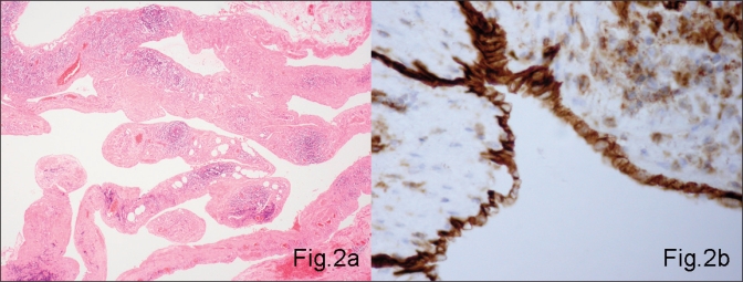
Fig. 2.
Histology of the cyst shows (A) lymphoid tissue and smooth muscle in its wall (hematoxylin and eosin stain, original magnification ×100), with (B) cuboid of flattened cells lining the cavity. Immunostaining with CD31 confirms their endothelial origin and hence the diagnosis of cystic lymphangioma (original magnification ×200).

