Figure 1.
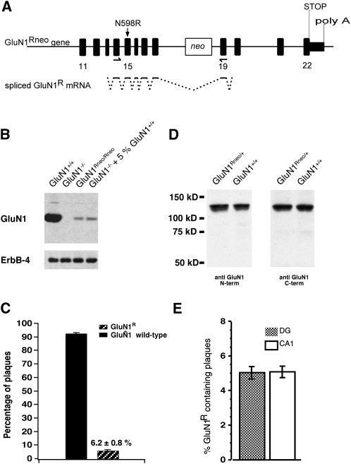
GluN1Rneo/+ mice express NMDAR GluN1R subunits at low levels. (A) Genomic organization of the GluN1Rneo allele with neo cassette in intron 18. Arrows indicate PCR primers for mRNA quantification plaque assay. Splicing that leads to the GluN1R hypomorph is indicated for the PCR fragment. (B) Western blot from P0 brain membranes, probed with antibody against GluN1 C terminus. The weak GluN1Rneo/Rneo signal is comparable to that obtained from GluN1−/− preparations spiked with 5% GluN1+/+ material (sample-loading control: ErbB-4). (C) Relative GluN1R mRNA abundance in forebrain of adult GluN1Rneo/+ mice (n = 5), determined by a plaque assay. (D) Western blot on adult brain homogenate. Antibodies against GluN1 N or C terminus revealed full-length GluN1 subunits and no truncated species in GluN1Rneo/+. (E) Relative GluN1R mRNA abundance of GluN1R transcripts from individual CA1 pyramidal neurons or DG granule cells (n = 7 cells, each region), determined by plaque assay.

