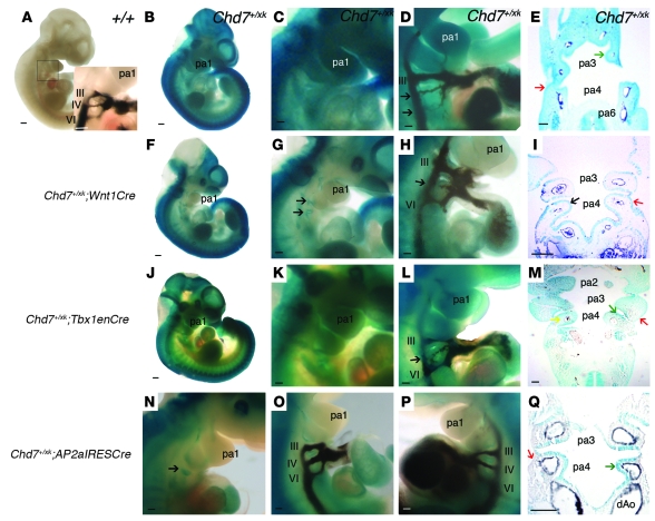
Figure 4. Effects of Cre-mediated rescue of Chd7 mutation on the arch artery phenotype at E10.5.
(A) WT embryo. Inset shows the normal PAA configuration. (B–E) Chd7+/xk embryos. (B) β-geo expression driven by the Chd7 promoter. (C) Magnified view of the pharyngeal region, showing ubiquitous expression. (D) Ink-injected Chd7+/xk embryo, revealing hypoplastic right fourth and sixth PAAs. (E) Reporter expression in all pharyngeal tissues (stronger in endoderm and ectoderm; arrows). Dark staining in arteries is India ink. (F–I) Chd7+/xk;Wnt1Cre embryos. (F) Loss of reporter expression in neural crest–derived structures. (G) Magnified view of the pharyngeal region, with trap retained in pharyngeal endoderm (arrows). (H) Hypoplastic fourth PAA (arrow). (I) Trap expression was lost in the pharyngeal mesenchyme, but remained in epithelia (arrows). (J–M) Chd7+/xk;Tbx1enCre embryos. Whole-mount embryo (J) and magnified views (K) of the pharyngeal region showed reduced X-gal staining. (L) Hypoplastic right fourth PAA (arrow). (M) Coronal section across pharyngeal region of a Chd7+/xk;Tbx1enCre embryo. Staining was reduced in core mesoderm, but was retained in ectoderm (red arrow). (N–Q) Chd7+/xk;AP2aIRESCre embryos. (N) Pharyngeal region, demonstrating loss of reporter staining in pharyngeal mesenchyme, but with staining present in pharyngeal endoderm (arrow). (O and P) Normal PAAs. (Q) Pharyngeal arch section showing loss of reporter activity in ectoderm and neural crest cell–derived mesenchyme, but maintained activity in pharyngeal endoderm (green arrow). dAo, dorsal aorta. Scale bars: 20 mm (A, B, F, and J); 100 μm (C–E, G–I, and K–Q).

