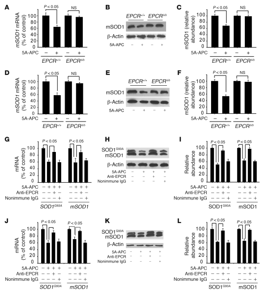
Figure 9. 5A-APC–mediated SOD1 downregulation in motor neurons and spinal cord endothelium requires endothelial EPCR.
(A–C) mSOD1 mRNA levels, determined by QPCR (A), and immunoblotting (B) and densitometry analysis (C) of mSOD1 protein, in laser-captured motor neurons from EPCRδ/δ and EPCR+/+ mice treated with saline or 100 μg/kg/d 5A-APC i.p. for 7 days. (D–F) mSOD1 mRNA levels (D), and immunoblotting (E) and densitometry (F) analysis of mSOD1 protein levels, in spinal cord microvessels isolated from EPCRδ/δ and EPCR+/+ mice treated as in A–C. (A–F) n = 3–4 per group. (G–I) SOD1G93A and mSOD1 mRNA levels (G), and immunoblotting (H) and densitometry (I) analysis of SOD1G93A and mSOD1 protein levels, in laser-captured spinal cord motor neurons of SOD1G93A mice treated with saline or 100 μg/kg/d 5A-APC i.p. for 7 days in the absence or presence of an EPCR blocking antibody (RCR-252) or nonimmune IgG infused through the femoral vein (40 μg/mouse) at day 1 and 3. (J–L) SOD1G93A and mSOD1 mRNA levels (J), and immunoblotting (K) and densitometry (L) analysis of SOD1G93A and mSOD1 protein levels, in spinal cord microvessels isolated from SOD1G93A mice treated as in G–I. (G–L) n = 3–5.

