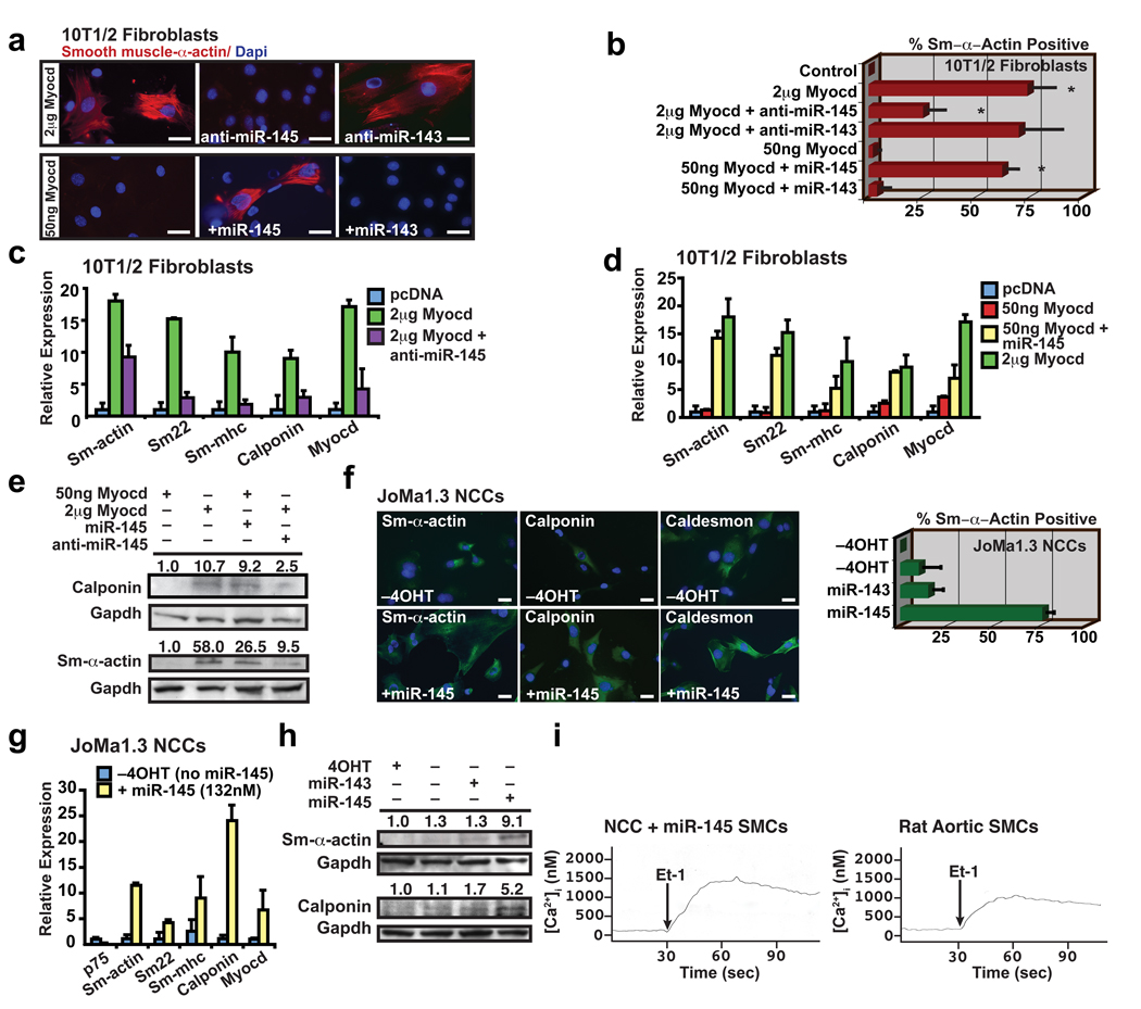
Figure 3. miR-145 directs vascular smooth muscle cell fate.
(a) Immunocytochemistry of 10T1/2 fibroblasts using smooth muscle (Sm) α-actin antibodies (red) under conditions indicated; nuclear stain, Dapi (blue). (b) Quantification of Sm-α-actin positive cells (n=6). (c) qPCR of Sm gene expression in fibroblasts transfected with Myocd with or without anti-miR-145 or (d) fibroblasts transfected with 50 ng Myocd with or without miR-145 (n=5). (e) Western blot of calponin and Sm-α-actin. (f) Immunocytochemistry of neural crest stem cells (Joma1.3 NCCs) with or without miR-145 using antibodies indicated (green); tamoxifen (4OHT) was removed to allow differentiation. Quantification of percent Sm-α-actin + cells relative to total Dapi+ nuclei (blue) (n=6). (g) qPCR of Sm gene expression in NCCs with miR-145 expression (n=5); p75 is a marker of undifferentiated neural crest cells. (h) Western blot of Sm-α-actin and calponin. (i) Calcium flux [Ca2+]i in SMCs derived from NCCs or rat aortic SMCs in response to endothelin-1 (Et-1) stimulation at 30 sec. Error bars indicating SD. *, p<0.05.

