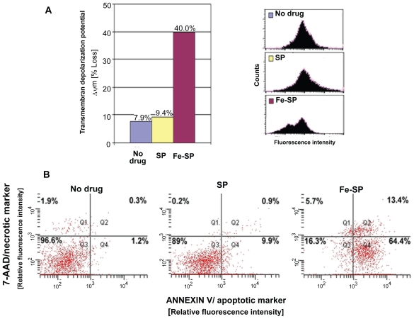
Figure 4.
Determination of mitochondrial membrane depolarization, apoptotic and necrotic SKOV-3 cells after Fe-SP treatment. (A) Mitochondrial membrane depolarization analysis. SKOV-3 cells were treated for 3 h with 3 μM Fe-SP or SP control, fixed and stained with DiOC18(3) as described (Materials and methods). Fluorescence of the single cell population was measured by flow cytometry (right panel) and the transmembrane depolarization potential of the single cell populations plotted (bar chart, left panel). Ten thousand cells were analyzed in each sample. (B) Apoptotic and necrotic cell population. SKOV-3 cells were treated with 1 μM Fe-SP or SP control for 24 h and floating and attached cells collected and combined. The quantification of apoptotic cells (Annexin V plasma membrane staining) and necrotic cells (7-AAD DNA staining) of SKOV-3 cells was carried out by flow cytometry as described (see Materials and methods). Ten thousand events were analyzed for each sample.

